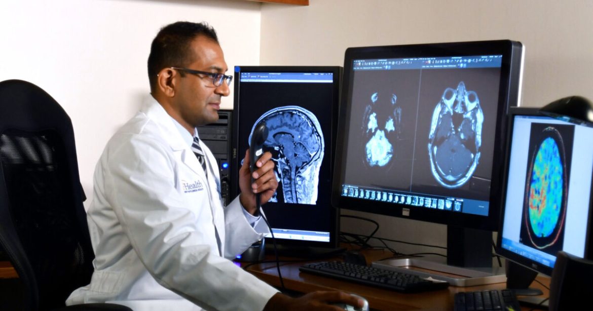
Neuroradiology plays a significant role in diagnosing and managing cerebrovascular disorders, offering precision and insight into conditions affecting blood vessels in the brain. Through advanced imaging techniques, it helps healthcare professionals detect issues early and develop targeted treatment plans. Here is how neuroradiology contributes to diagnosis and treatment:
What Are Cerebrovascular Disorders?
Cerebrovascular disorders are conditions that affect the blood vessels in the brain. These problems can disrupt blood flow and, in severe cases, cause a stroke. The group of conditions includes aneurysms, artery blockages, and vascular malformations. When blood flow is interrupted or altered, it can directly impair brain function and sometimes cause permanent damage. Detecting these issues early is needed to prevent complications.
Symptoms vary widely but often include headaches, dizziness, numbness, or difficulty speaking. Quick detection may improve the chances of successful treatment. Neuroradiology plays a fundamental role by offering detailed scans that reveal the brain’s structure and function.
How Does Imaging Work?
Neuroradiologists use advanced imaging techniques to better understand brain problems. They rely on methods like CT scans, MRI scans, and angiography, each providing unique information about brain structure and blood flow.
- CT scans are typically the first choice in emergencies because they quickly show bleeding or other abnormalities in the brain.
- MRI scans offer detailed images of soft tissues, making them especially useful for detecting strokes and small structural changes.
- Angiography concentrates on blood vessels, helping identify blockages, aneurysms, or malformations accurately.
These imaging approaches help medical teams understand the type and severity of cerebrovascular disorders, guiding better treatment decisions.
How Are Treatments Guided?
Once a diagnosis is made, neuroradiology continues to be a core element in treatment. It guides procedures and monitors progress. These procedures often involve threading a catheter through blood vessels to reach the brain directly. This allows for treatments like placing stents or coilings of aneurysms. Before the procedure, imaging provides a roadmap to visualize the path. During the procedure, real-time imaging improves accuracy. Afterward, follow-up scans check the effectiveness of the treatment and determine if additional therapy is needed.
What Happens After Treatment?
Recovery and monitoring are key parts of cerebrovascular care. Neuroradiology remains fundamental even after treatment ends, as it is used to assess recovery and prevent relapse. Regular follow-up scans help healthcare providers monitor healing and detect any recurring problems. After aneurysm surgery, periodic imaging checks ensure no new issues have developed.
This ongoing monitoring lowers risks and supports long-term brain health. Beyond imaging, many patients take part in rehabilitation programs aimed at regaining physical functions. Physicians use findings to tailor these programs to the patient’s specific needs, so that their recovery is appropriate for their condition.
Why Is Neuroradiology Significant?
Neuroradiology transforms cerebrovascular care. Its ability to visualize the brain’s complex network of blood vessels empowers medical teams to diagnose, treat, and monitor like never before. With technologies such as CT, MRI, and angiography, patients have access to life-saving interventions that might not have been possible just a few decades ago. Effective use of these tools has brought remarkable improvements in patient survival rates, outcomes, and quality of life. Schedule an appointment with a radiologist near you.
